| GENERAL
Introduction
General Overview
Clinical Services
Research
Teaching
PhDs & Projects
Personnel
IMAGE PAGES
Visual overview
CT, MRI Images
3D Ultrasound
Fetal Studies
Surface Scanner
UCL Hospitals
|
MEDICAL IMAGING GROUP
UCL and UCL Hospital
A Cranioplasty plate is used to fill a defect in the skull, which may
arise from trauma, surgery or other cause. We have a great deal of experience
with Titanium cranioplasty plates. There are many reasons for preferring
these to other materials (Ref 1) and we have a current project assessing
these. Although we could probably provide cranioplasty plates in other
materials using our automated procedure, our standard is currently Titanium.
We take a 3D image (set of CT slices) of the required segment of skull,
which can be either scanned here or on any scanner which can output data
in a known format (we know a good many). We then reconstruct the 3D image,
using our own 3D workstation.
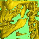
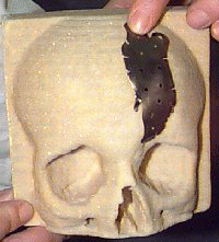
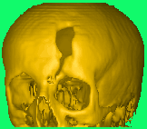 Skull
trauma Skull
trauma
The prosthesis is then planned. This process may include using a mirrored
section of skull on the opposite side of the skull, to obtain a contour
where large segments are missing. From the plan a mould is prepared using
computer controlled milling. This mould is thus prepared and shaped directly
from the imaged skull bone surface and will match the surface accurately.
The plate is then pressed directly from this mould.
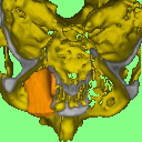
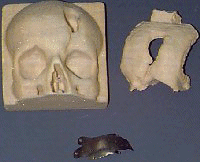 Milled
model, cast model, pressed titanium Milled
model, cast model, pressed titanium
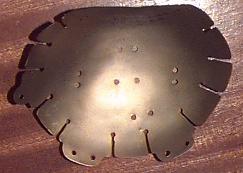
In this way a plate can be made which accurately fits the skull at surgery,
including the minimum number of screw holes or tabs. The 3D imaging provides
us with all the required information about bone thickness etc, required
to place a minimum number of these mounting screws. Plates can be prepared
prior to surgery, so that there is never a need for a second operation
to fit the cranioplasty plate. We then supply the anodised Titanium plate,
together with Ti screws, ready for insertion.
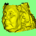
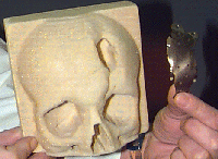 Cranio in place Cranio in place
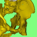
 Model & cranioplasty plate Model & cranioplasty plate
 Example of
a larger plate We are able to accept 3D images in a variety of data formats, from
different scanners, on different media, including the Internet.
It is unfortunately not possible, at this moment in time, to transfer
the finished plates over the internet. Should such material hyperlinks
become available, we will be the first to make use of them. Example of
a larger plate We are able to accept 3D images in a variety of data formats, from
different scanners, on different media, including the Internet.
It is unfortunately not possible, at this moment in time, to transfer
the finished plates over the internet. Should such material hyperlinks
become available, we will be the first to make use of them.
This work is directed by our professional prosthetist, in close collaboration
with the Dept. of Maxillo-Facial Surgery of UCL/UCLH.
These techniques can also be used to create surgical
models for a variety of purposes including visual aids to complex
surgery in Maxillo- facial, orthopaedics and other areas.
Other Clinical Applications:
|
 |