| Read
Display
Info if you have difficulty browsing these graphics-orientated pages. |
To view a movie, click on an image with a
[GIF] or [AVI] link) |
|
|
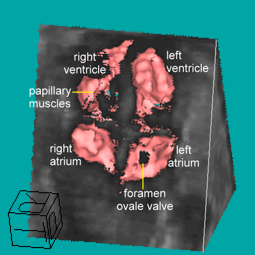
| Cardiac motion gated 3D image of a 22-week fetal heart
in utero. Fine structures such as the foramen ovale valve and papillary
muscle are depicted. [GIF 286KB]
[AVI
387 KB] |
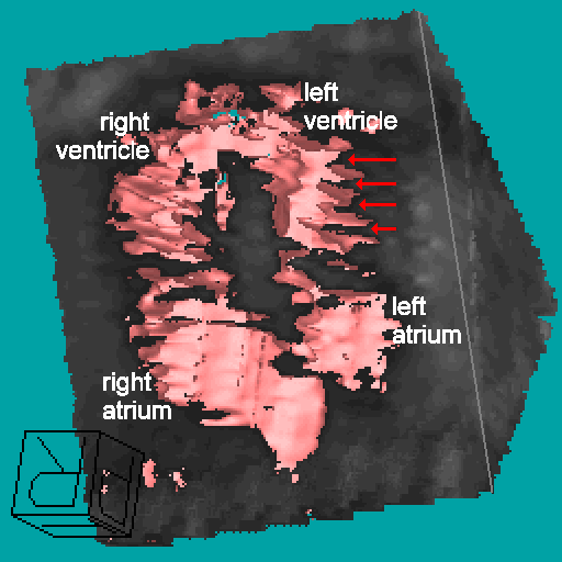
Before gating, fine structures are degraded by motion
artefacts (red arrows). [GIF 311KB] |
|
|
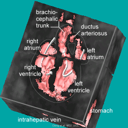
| Another cut of the thorax (and abdomen) shows
an end-systolic gated view of the four chambers and great vessels. [GIF
340KB] |
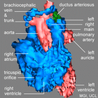
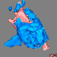
The same fetal heart in a negative surface
display (i.e., cavities shown in solid) showing the spatial relationships
of the cardiovascular structures. End-systolic gated. A shorter movie [GIF
784KB] |
|
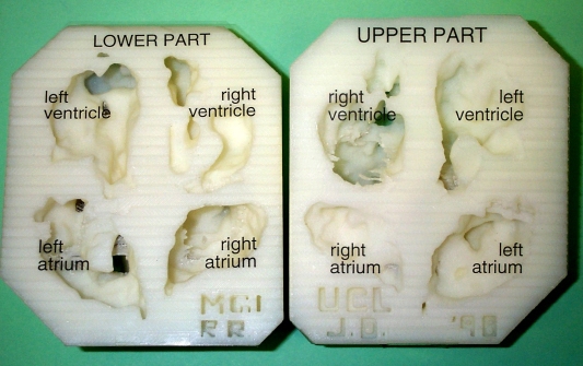
| This is a pair of cardiac models prototyped
from the 3D ultrasound data set of the same live fetal heart (above).
More
about PROTOTYPING with digital image data sets from this group. |

![[GIF 784KB]](hrt3sysn5-200s.gif)