| GENERAL
Introduction
General Overview
Clinical Services
Research
Teaching
PhDs & Projects
Personnel
IMAGE PAGES
Visual overview
CT, MRI Images
3D Ultrasound
Fetal Studies
Surface Scanner
UCL Hospitals
|
MEDICAL IMAGING GROUP
UCL and UCL Hospital
Surgical Model Manufacturing Services
We can manufacture models of most structures which are of interest
to surgeons. These are made directly from 3D surfaces which may be derived
from CT, MRI or Ultrasound using our own 3d imaging
system. We use a computer-controlled milling process to manufacture
models from blocks of polyurethane foam. The usual application is in complex
bony formations (from CT), for which these models are found to be extremely
useful when complex fractures are being surgically repaired.
These models are made in house, and can be prepared quickly if required.
We can also make models using stereolithography (using an outside service)
if required, but this method is currently rather more expensive.
 An
intermediate cost alternative is fused deposition modelling. We now have
our own Stratasys FDM 3000 system and can manufacture high quality models
in ABS plastic in house. This will considerably shorten the time between receiving patient data and having a model ready. An
intermediate cost alternative is fused deposition modelling. We now have
our own Stratasys FDM 3000 system and can manufacture high quality models
in ABS plastic in house. This will considerably shorten the time between receiving patient data and having a model ready.
We are able to accept 3D images in a variety of data formats, from
different scanners, on different media, including the Internet.
The completed models can then be mailed direct to the customer if required.
|
|
|
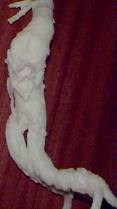
| The first ABS model made on our new FDM 3000 rapid prototyper.
Slippage of stent graft after endoluminal abdominal aneurysm repair:
note single right renal artery with branch. (more images & movie here)
|
This is an early example of the use of rapid prototyping . A polyurethane
foam model milled directly on our CNC milling machine in 1990.This image
also shows theTitanium cranioplasty plate
custom made to repair the defect. |
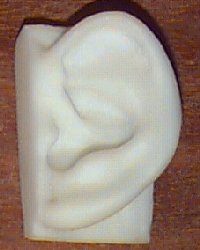
An ABS model of an ear used for prosthesis design and manufacture |
|
|
|
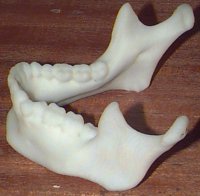
An ABS model of a mandible |
Another early example of the clinical use of RP. This plaster model was made in 1991 by milling two negative mould parts in polyurethane foam and filling the mould with plaster of paris. It shows a compressive fracture of the top of the tibia (knee-joint). This model was a valuable aid at surgery. |
|
Other Applications:
For more information please email Robin Richards, robin@medphys.ucl.ac.uk
|
 |