| GENERAL
Introduction
General Overview
Clinical Services
Research
Teaching
PhDs & Projects
Personnel
IMAGE PAGES
Visual overview
CT, MRI Images
3D Ultrasound
Fetal Studies
Surface Scanner
UCL Hospitals
|
MEDICAL IMAGING GROUP
UCL and UCL Hospital
3D CT Imaging
We have been developing 3D imaging techniques for about 15 years, using
hardware and software developed here at UCL
and UCL Hospitals.
This page focuses on some of the methods which have proved of considerable
value and interest using X-ray CT (Computed Tomography). These techniques were made possible by the use of a 3D
workstation with facilities for rapid processing and a range of sophisticated
interactive facilities. First some hard tissue images (bone) then soft tissues.
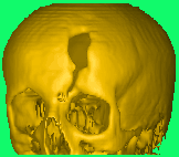 |
This image shows a skull requiring a cranioplasty to cover the defect.
We manufacture anatomical models and titanium
cranioplasty plates as one of our routine NHS
services (images can come via the internet but the manufactured plates
still need to be sent by post). |
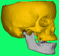 |
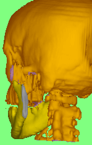
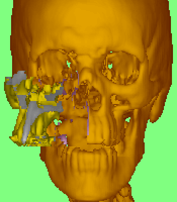
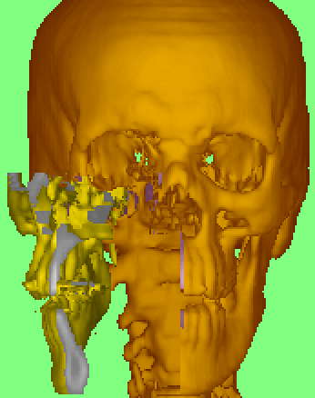
This image shows how image 'surgery' can be applied, in this case separating
and moving the mandible. |
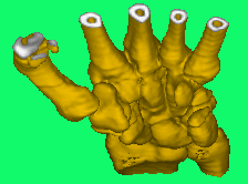
Wrist and fingers
 |
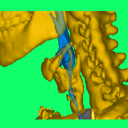
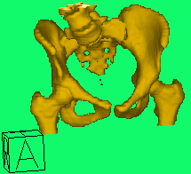
Pelvis
 |
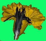 |
Hard tissue surfaces are usually straightforward to render in 3D. However
for soft tissues we need to be able to see inside. This study of a
carnation which had been given an iodine contrast agent shows the surface
rendered in gold and the internal structures as a normal CT greyscale where
the (imaging) cuts have been applied.
( No abnormality was found and the subject was asymptomatic after 24 hours.) |
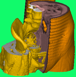 |
This image shows a number of different tissue types (soft tissue, muscle
and bone) in a single image by selection of different thresholds and different
cuts through the data. |
The following mpeg videos show proposed strategies for surgical access
to the deep pharyngeal region.
|
 |