UCL DEPARTMENT OF MEDICAL PHYSICS AND BIOENGINEERING
FACULTY OF ENGINEERING SCIENCES
UCL logoInformation Systems banner

GENERAL
Introduction
General Overview
Clinical Services
Research
Teaching
PhDs & Projects
Personnel

IMAGE PAGES
Visual overview
CT, MRI Images
3D Ultrasound
Fetal Studies
Surface Scanner

UCL Hospitals

MEDICAL IMAGING GROUP
UCL and UCL Hospital

Overview of 3D Imaging

We have been developing 3D imaging techniques for about 15 years, using hardware and software developed here at UCL and UCL Hospitals. This is a visual overview. For more text and technical details see our General Overview page.

These techniques were made possible by the use of a 3D workstation with facilities for rapid processing and a range of sophisticated interactive facilities.

A 3D (medical) image is the data set recorded from a solid volume of tissue, rather than the views and projections used to represent it. Views are always 2 dimensional (because screens and retinas are) and there is usually hugely more information in a 3D diagnostic image than can be represented on a 2D screen even with clever volume rendering or colour coding. The following gives a brief illustration of some of the methods and uses.


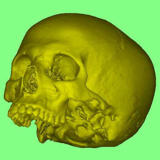
Fig. 1. A skull image

Skull
This is a 3D rendered view of a high quality CT image of a dry skull. Most of the required information is seen on this view; or on rotating; or on extracting detail.
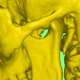
Skull detail.

However if we scan a complete head, there is information within the skull which can be seen by making cuts.
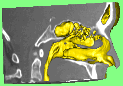
Skull with defect MPR.

Cutaway views can be produced in many different ways, as shown in this image of the pelvis/sacrum.
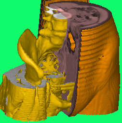

Pelvis/sacrum.

The following image shows an (abnormal) airway highlighted within the bony structures of the head and neck. Only these 2 components are visible.
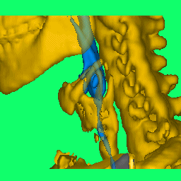

Airway.


University College London - Malet Place Engineering Building - Gower Street - London - WC1E 6BT - Telephone: +44 (0)20 7679 0200 - Copyright © 1999-2005 UCL


Search by Google