GENERAL
Introduction
General Overview
Clinical Services
Research
Teaching
PhDs & Projects
Personnel
IMAGE PAGES
Visual overview
CT, MRI Images
3D Ultrasound
Fetal Studies
Surface Scanner
UCL Hospitals
|
MEDICAL IMAGING GROUP
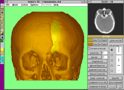
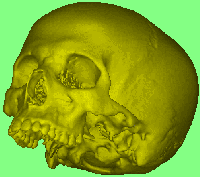
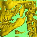
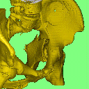
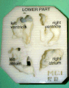
UCL and UCL Hospital
3D Imaging Workstation
The MGI Workstation operates on 3D Image data derived from a variety
of sources of section scan data or from Laser surface scans. Developed
for Maxillo-facial surgery, this has found many applications in orthopaedic
and other surgery. The user can use a variety of cutting, repositioning
and joining procedures on the 3D data, which will have been acquired from
CT, MRI or even 3D ultrasound. Surface views of the manipulated image can
be viewed from any direction; sectional images can be reconstructed along
any plane, allowing full assessment of the spatial image structure modified
by the user. This device is an essential part of most of our 3D work.
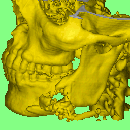
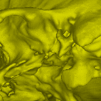
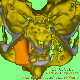
The original workstation used a network of Transputers as the processing
engine and achieved very fast rates for reconstruction of various types
of views, always operating on the original data slices, rather than using
geometric reduction such as polygons. Surface views using ray-tracing;
interactive multi-planar reformating; MIP and summed voxel projections;
cut surfaces; image surgery, involving cutting and joining, model
making and various measurement facilities are available. Versions of it have been installed in a number of hospitals in the UK and
elsewhere.
A new much more powerful model is now being developed using modern and
lower cost technology (PC). This is now available commercially.
 |
|
Some example images
Other Clinical Applications:
For more details of the workstation and its software contact Robin Richards. Email: robin@medphys.ucl.ac.uk
|
 |