This patient has had customised titanium implants fitted
to restore a more natural contour around the eyes.

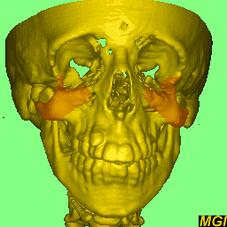
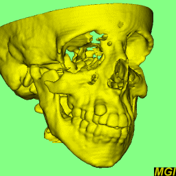
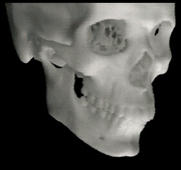
The image above shows the patient's deficient malar (top) and the normal
anatomy (bottom). The basic implant shape was copied from the normal skull
and a rapid prototype model of the patient was manufactured to ensure that
the shaped plates would fit correctly on the patient.
|
|
|
|
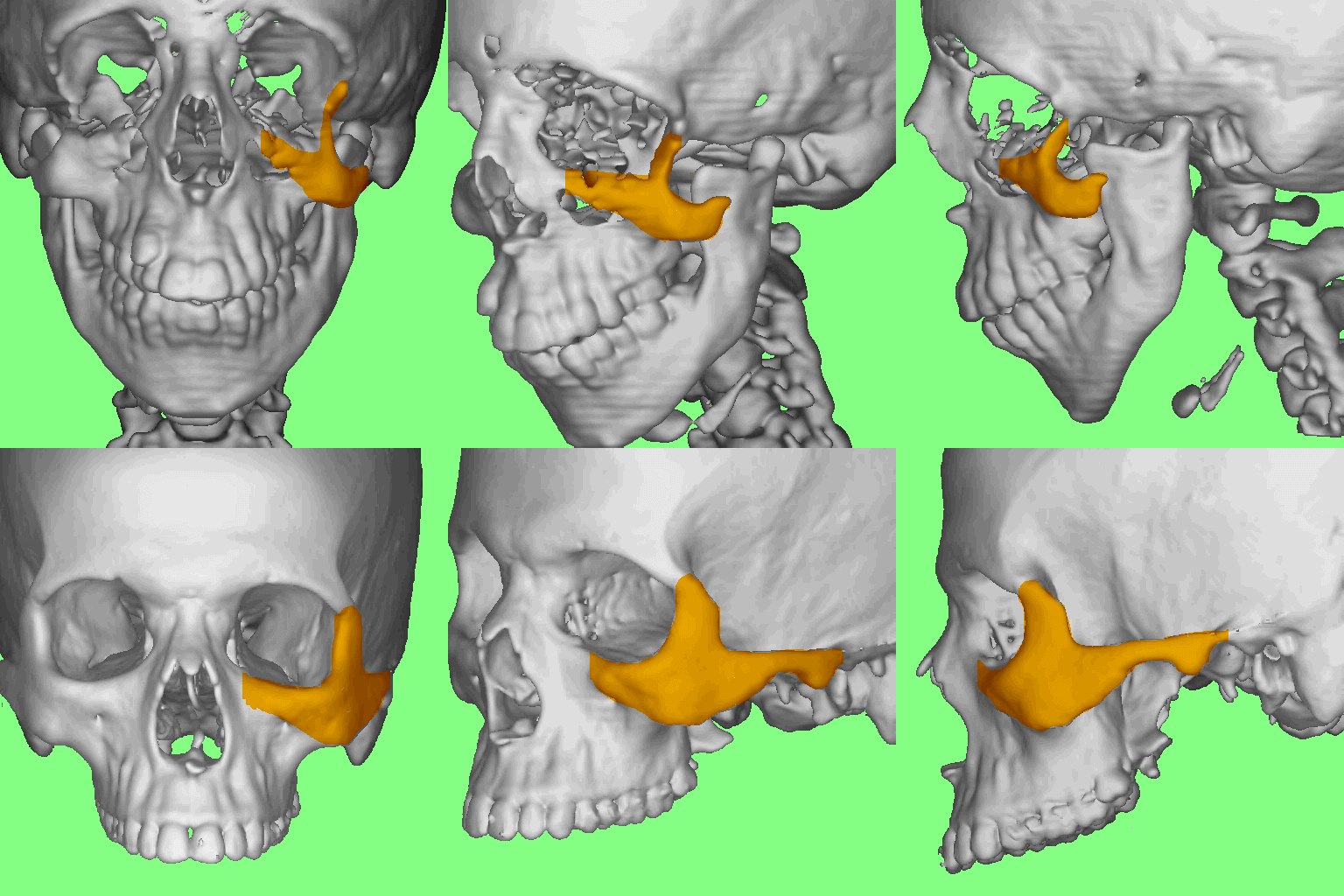
| Image from the CT scans showing deficient growth in the malar
region (highlighted) (mpg movie 780Kbytes) |
Stereolithography file generated from the CT scans for manufacture
of a physical model ( mpg movie 780Kbytes) |
|
 |
 |
|
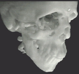
ABS plastic model of normal anatomy
(mpg movie 1Mbyte)
(mov movie 1Mbyte) |
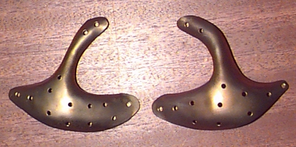
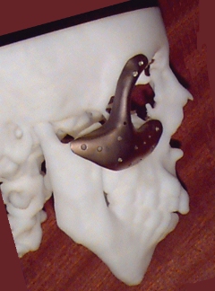
The titanium plates ready for implanting |
|
|
|
 |
 |
 |
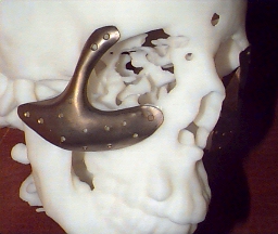
The ABS plastic model with the titanium implants mounted on it
(mpg movie 917Kbytes)
(mov movie 901Kbytes)
(avi movie 333Kbytes) |
|
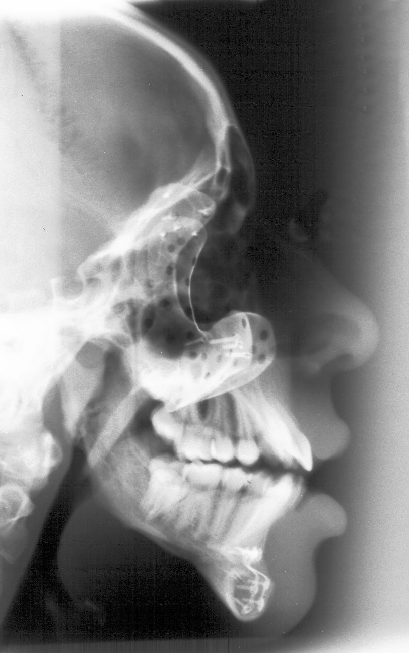
Lateral X-ray of patient showing the implanted plates in place |
Models of the patient with and without the implants and a model of a
reference skull are on display in the Digitopolis
section of the Wellcome
Wing of the Science Museum