THE COST
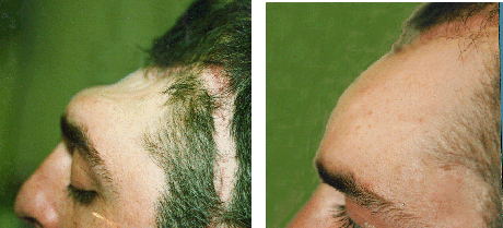
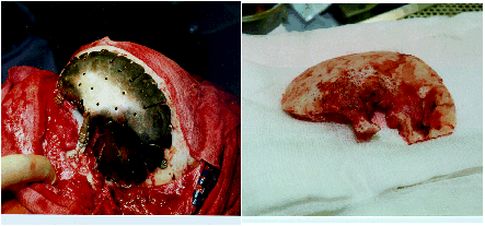
Views of patient before and after insertion of cranioplasty plate
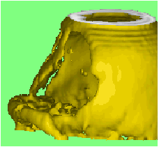
3D computerised image of the defect and a finished plate
The Cranioplasty Unit at UCLH is able to
provide highly accurate titanium plates, using a computerised technique
based on axial CT scans of the skull and defect. We have been providing
this service since 1990 and have a great deal of experience with the design
and manufacture of titanium cranioplasty plates.
A 3D image of the skull is generated from
the CT scans, acquired either here or from any scanner that is compatible
with our 3D workstation, or sent across the Internet. The scanners we support
are listed below and we can arrange to add further scanners to this list
if necessary.
The 3D image provides us with all the required
information about surrounding bone thickness and most suitable attachment
sites. Mirror imaging is often used to restore contour for bi-lateral defects.
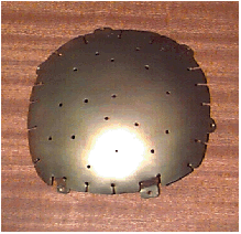
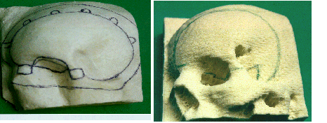
A life-size model of the defect is then automatically produced on a computer
numerically contolled (CNC) mill. A mould is prepared from the model incorporating
the design of the plate, and the plate is then constructed by pressure
forming sheet titanium from the mould of the recontoured skull1.
Plates can be prepared for the repair of
pre-existing defects or prior to surgery eliminating the need for a second
operation to fit the plate. We supply the anodised titanium plate, together
with the titanium screws, ready for insertion.
1. Joffe JM, McDermott PJC, Linney AD, Mosse
CA and Harris M. Computer Generated Titanium Cranioplasty: Report of a
new technique for repairing skull defects. British Journal of Neurosurgery
1992; 6: 343-350.
THE SERVICE
When using this service, we would like you
to provide us with the following:
A referral letter to the Cranioplasty Unit
indicating your requirements. This should include the patient's name and
age and when the plate is required. Also, it would be most helpful if you
could provide us with clinical information if the site of the defect could
affect the design or the attachment features of the plate. (Eg. elimination
of frontal sinuses, or when a defect is in a position involving an area
known to have thin bone, such as within the temporal fossa, or if there
is to be any further removal of bone at time of fitting the plate.) Plates
are usually made within 3 weeks, but for urgent cases this could be a few
days. Please enclose an order number or arrange for your Finance Department
to raise an order. For private patients the invoice will normally be sent
directly to the Consultant requesting the plate.
A tape or discs containing CT scan data of
the skull. This should be sent to the Cranioplasty Unit at the address
below. The recommended protocol for CT scanning is stated below. Please
give your radiographers this information.
CT SCAN PROTOCOL
Position the patient's head in the headrest
so that as many slices as possible pass through the defect. Once positioned
make sure the patient does not move during the scanning session to ensure
good registration.
Scan the patient with contiguous sequential
slices to cover the area of the defect and at least 2cm of the surrounding
tissue. We recommend a slice separation of no greater than 3mm through
the defect. If you have a helical scanner then 3mm or 5mm spacing could
be used with the slices re-interpolated to 1.5mm or 2.5mm. Keep the field
of view constant, do not alter the couch height, and if possible use no
gantry tilt. Standard reconstruction algorithms should be used.
The processed image data should then be stored
on one of the listed archive media, or transferred across a network. Send
the tape or discs to the Cranioplasty Unit at the address below. It is
helpful initially if you could also send a film containing the scanogram
and a few of the scans. This verifies scaling, any gantry tilt and orientation.
Please contact us if you have any queries
about scanning or if your scanner is not listed here before scanning the
patient.
THE COST
PLATES
We have 4 categories of the plates depending
on the size and the complexity: small, medium, large and complex. Please
call for the current prices.
An estimate can be given after seeing the CT
scans.
SCREWS
We specify the size of the bone screws to be
used with the plates: they are 2mm in diameter, 6mm/4mm in length. You can
use your own choice of titanium screws, or we can supply the Stryker bone
screws for you with extra costs.
The construction of a primary pre-operative cranioplasty plate