Photoacoustic-photothermal
probe
Sensor head |
Performance | Applications | References |
Contact
Pulsed photoacoustic and
photothermal techniques are investigative methods in which short
sub-ablation threshold excitation laser pulses are absorbed in a target
absorber producing both acoustic (thermoelastic) and thermal waves.
These waves act as carriers of information relating to the optical,
acoustic and thermal properties of the target absorber and can be used
to describe its constituents and structure. Applications include the
characterisation of biological tissue and non-destructive testing of
materials and structures. Whilst photoacoustic and photothermal
techniques provide an inherently powerful means of characterising a
target, their practical implementation can be problematic using
conventional acoustic and thermal detection methods. This is
particularly so when it is required that the generation and detection of
the photoacoustic or photothermal signals take place on the same side of
the target as is generally required for the in situ
characterisation of biological tissues. In such cases the
acoustic/thermal detector should be transparent so that it can be
aligned coaxially with the excitation laser beam thus excluding most
conventional piezoelectric and pyroelectric contact transducer
configurations. Biomedical implementations also often require a
miniature, flexible probe type format for minimally invasive use such as
insertion via a biopsy needle or endoscope and this too represents a
substantial challenge using existing methods. A miniature all-optical
probe [1,6]
that employs a transparent acoustically and thermally sensitive Fabry
Perot sensor for making photoacoustic and photothermal measurements
simultaneously has been developed that offers a potential solution to
these limitations.
Back to top
Sensor head
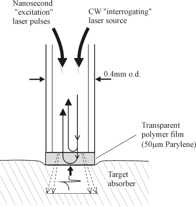
Figure 1 Schematic of the
sensor head probe.
Principles of operation: A schematic of the sensor head probe is
shown in figure 1. A 380µm core diameter multimode optical fibre with a
transparent Fabry Perot polymer film sensor mounted at its distal end is
placed in contact with the target absorber (figure 1). Nanosecond, sub-millijoule
optical pulses from a Q switched Nd:YAG laser are launched into
the fibre, transmitted through the sensor and absorbed in the target
producing thermal waves with a typical duration of the order of a few
hundred milliseconds. In addition, rapid thermal expansion occurs
generating ultrasonic thermoelastic waves with a typical duration of
several hundred nanoseconds. Both thermal and thermoelastic waves are
detected by the sensor at the tip of the fibre. The sensor itself
comprises a transparent 50μm thick
polymer film. This can either be deposited under vacuum directly on to
the cleaved end of the fibre using the Parylene process or a discrete
polymer film such as PET mounted at the fibre can be used. When
illuminated by light launched into the fibre from a CW low power
tuneable laser source, the polymer film acts as a low finesse Fabry
Perot interferometer with the refractive index mismatches on either side
of the film providing the mirrors of the interferometer. An incident
thermal or thermoelastic wave changes the optical thickness of the film
and hence the optical phase difference between the Fresnel reflections
from either side of the film. This produces a corresponding intensity
modulation in the light reflected from the sensing film which is then
detected by a photodiode. Linear operation is achieved for small
measurand-induced phase shifts by tuning the wavelength of the laser
source so that the interferometer phase bias is set to the optimum
quadrature point.
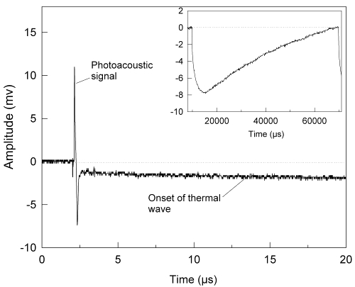
Figure 2 Sensor output (50mm
water-backed PET film) in response to photoacoustic and photothermal signals generated in an ink-Tris absorber (ma=70cm-1).
Inset shows complete thermal signal over expanded timescale.
Fluence: 0.27mJ/mm2,
pulse duration: 5ns, repetition rate: 16 Hz. [1]
Experimental
set-up and procedure: Figure 2 demonstrates the dual sensing
ability of the system showing the photoacoustic and photothermal signals
generated in an ink/Tris solution of absorption coefficient µa=70cm-1
over different timescales. Figure 2 shows the short duration
(400ns) photoacoustic signals. The step decrease, immediately following
the initial thermoelastic wave is due to the initial heating of the
target by the laser pulse and can be regarded as the onset of the rising
edge of the thermal wave. This very slow increase in the thermal signal
can be seen more clearly in the inset which shows an expanded timescale
graph.
Back to top
Performance
The acoustic system sensitivity
was obtained by comparing the sensor output to a calibrated 25MHz PVDF
membrane hydrophone and was found to be 140 mV/MPa with an acoustic
noise floor of 2 kPa over a 25 MHz measurement bandwidth and 30
averages. The dc thermal system sensitivity was established by placing
the sensor head in a water bath and recording the sensor output as the
temperature was varied. A calibrated thermocouple placed immediately
adjacent to the sensor head was used as a reference. The dc thermal
system sensitivity was found to be 32 mV/ºC with a thermal
noise floor of 6.3 x 10⁻³°C,
also over a 25 MHz measurement bandwidth and 30 averages. By varying the
temperature over 25°C, it was possible to observe a maximum
and minimum of the interferometer transfer function (indicating a phase
shift of π rad) giving a temperature (phase) sensitivity of
0.13rad/°C.
Back to top
Applications
In vivo
measurement of tissue optical properties
Analysis of the amplitude and temporal characteristics of the
photoacoustic and photothermal signals can yield the optical properties
of the target tissue and can be used to discriminate between different
tissue types. For example, photoacoustic spectroscopy [7]
has been used to characterise arterial tissues based upon the strong
preferential absorption in atheroma at visible wavelengths and we are
currently investigating the use of photothermal
methods for the detection of cancers [6].
The flexibility and small size of the probe offers a means of
implementing these techniques in vivo in a minimally invasive
form such as via a biopsy needle, endoscope or catheter. Additionally
the use of polymer film deposition techniques enables the sensor to be
batch fabricated at low unit cost for disposable use to avoid cross
infection.
Laser
ablation studies The probe has application in fundamental studies
of laser ablation where there is a need to measure the acoustic and
thermal transients generated and relate them to the degree of disruption
produced in the target. For example, in biomedical laser ablation
processes such as laser angioplasty the acoustic transients generated
can produce damage to the vessel wall beyond the ablation site and it
has been suggested that the resulting increased trauma to the vessel may
play a role in stimulating restenosis. It would be useful to quantify
such photomechanical effects and additionally any photothermal response
by the direct measurement in vivo of the laser-induced acoustic
and thermal transients and relate them to the observed physiological
response.
Non-destructive testing (NDT)
- The probe may also act as a miniature all-optical laser ultrasound
transmitter and receiver for laser NDT/ND evaluation applications (e.g.
flaw detection in engineering structures) that require an inexpensive
integrated probe capable of making same-side coaxial measurements. The
flexible nature of the optical fibre downlead and its very small
diameter (0.25mm) provides a means of making measurements in locations
that are difficult to access such as pipes or small cavities. The
inexpensive nature of fabrication allows the probe to be considered an
inexpensive consumable that can be used in hostile environments where
there is a risk of damaging expensive piezoelectric transducers. Since
the probe is electrically inert it can be safely deployed in
flammable/explosive environments where there is a risk of ignition from
a electrical sources and used in electromagnetically noisy environments.
Back to top
|