1. Context
I am Yassine
Bouchareb, clinical scientist in image fusion working full time on
the multi-modality medical image fusion project. This project is run within
the Department of Medical Physics
and Bioengineering of University College of London Hospitals by Prof.
Roland Blackwell (Head of the department) and the Institute
of Nuclear Medicine, University College London by Prof. Peter Ell (Director
of the institute).
Several Radiologists are involved in this project: Liz Prvulovich,
consultant physician in Nuclear Medicine, Nicholas Hyde a maxillofacial
surgeon at the Maxillo Facial Unit and Rolf Jager, consultant Neuroradiologist
at University College London Hospitals Trust and Reader in Neuroradiology
at the Institute of Neurology,
National Hospital, Queen Square.
2. Objective
The first aim of the project is to implement an image fusion application
for ontological studies using images acquired using the GE "Advanced" full
ring PET scanner in the Department of Nuclear Medicine and images acquired
using the Large Bore Picker "Acqsim" CT scanner used for simulation in
the Radiotherapy Department. The fusion application will be delivered for
clinical use for radiotherapy treatment planning. Further application for
MRI-PET fusion are being investigated.
3. Progress & Results
The Image Fusion Project is based on the use of the rigid body multi-modality
image fusion software developed at Guys Hospital for research purposes.
This fusion technique is voxel intensity based and uses mutual information
as a similarity measure for registering images. This software is actually
installed and running on the sun workstation "chaabi" dedicated to this
project. The project was started by working on CT and PET data. Some preliminary
results (click
here to see the presentation) for the chest scans CT/PET were presented
at the UK PET physics group
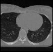
on 10th May (see figure 1).
Figure1. CT axial slice, a
PETemission slice, superimposition of CT/PET images
before registration
and the fused images from left to right.
Six datasets for Chest patients have been processed
so far, and there is still great interest in recruiting more Brain, Chest
and Head & Neck studies from Radiotherapy to increase the range of
clinical applications covered, and a more accurate evaluation of our fusion
technique.
Recently 12 Head & Neck patients with both
MRI and PET scans have been processed in order to test the ability of the
fusion application to show how accurate are these fused images (see figure
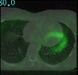
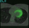
2a and 2b).
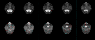
Figure2a. MRI T1 (left) and
PET (right) images from a Head and Neck patient.
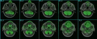
Figure2b. MRI/PET fused images
from images above.
The fusion results for these Head and Neck patients
are interesting from a clinical point of view, however, these results show
some limitations of the actual MRI acquisition protocol in addition to
the type of deformations that are likely to be present in this region.
To overcome these limitations, the practical
ways to proceed for the MR imaging protocol for Head and Neck studies were
discussed in the group. A specific MRI acquisition protocol for image fusion
will be considered for the future tumour patients candidates for the fusion
process. This protocol is currently under test.
The MRI protocol should include T1 transverse
slices spaced at 3 mm through the region of interest (bottom of the brain
to the top of shoulders). The time implications of including the T1 scan
in the protocol will be studied as well.
In the future, the use of MR images acquired
with an appropriate acquisition protocol for image fusion is expected to
increase further the value of this fusion technique.
4. The Image Fusion Group