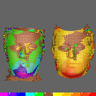
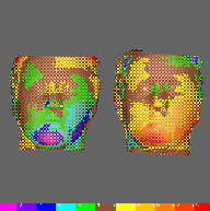
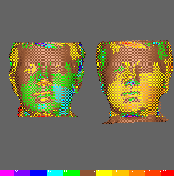
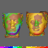
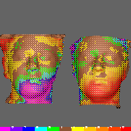
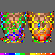
Right of page: Coded Difference image for this pair.
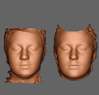
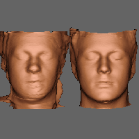
Female Age 17+
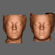
Craniofacial Microsomia/ Control
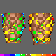
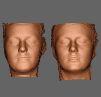
Registered Scans
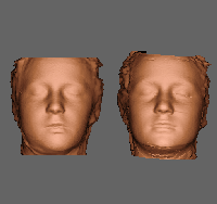
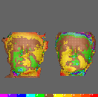
Female Age 13-16
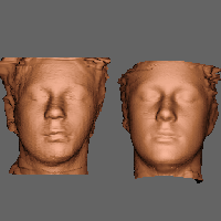
Craniofacial Microsomia/ Control
Registered Scans
Female Age 9-12
Craniofacial Microsomia/ Control
Registered Scans
Female Age 4-8
Craniofacial Microsomia/Control
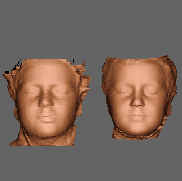
Registered Scans
Male Age 4-8
Craniofacial Microsomia/ Control
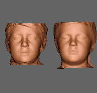
Registered Scans
Male Age 9-12
Craniofacial Microsomia/ Control
Registered Scans
Male Age 13-16
Craniofacial Microsomia/ Control
Registered Scans
Male Age 17+
Craniofacial Microsomia/ Control
Registered Scans
**** End of PhD Poster. ****
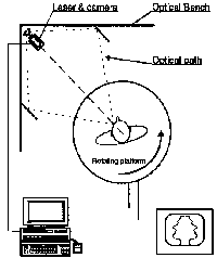
For more details of laser surface scanning and its application or to get in touch with us, please see our main pages:
M G I Group, UCL/UCL Hospitals.
 Go to Research Page
 Return to Home Page
Poster created by Tricia Goodwin. , E-mail: pmg@medphys.ucl.ac.uk,
Web page version edited by John Gardener, E-mail: jeg@medphys.ucl.ac.uk
Last update 22/1/97.
 Back towards the Home Page