Intravascular ultrasound (IVUS) is recorded using a very small probe (1mm x 3mm) which rotates inside a water-filled catheter. This conventionally produces 2D images which are approximately cross-sections of the vessel.
A 3D image is recorded by pulling back the catheter along the vessel axis, recording its position and grabbing a set of 2D images (typically 80-100). This is then reconstructed assuming linear motion, and viewed on one of our workstations, which allows cutting away; multi-planar reformatting (MPR) and other views.
The following gives a quick visual outline of some of the work we have done.
<
Fig. 1.


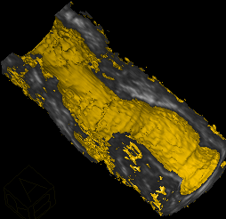
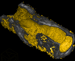
Two views of an iliac artery (in-vivo), recorded using intravascular ultrasound. The recorded 3D image has been cut in half to allow visualisation of the internal lumen. A small band of plaque may be seen in a spiral around the vessel above the bifurcation. The resolution of this image @20 Mhz is about .2 mm.
To see this as a video click here:
Iliac IVUS Video
There is more material to come - watch this space!