The following gives a quick visual outline of some of the work we have done so far.
Fig . 1.
A fetal face at about 16 weeks. The left hand is in front of the mouth.
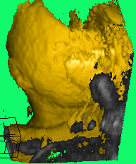
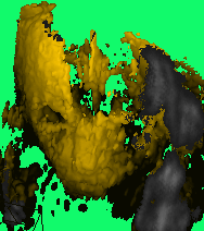
Fig . 2.
A whole fetus at about 18 weeks showing face, trunk arms and legs. The left arm (at right) is partly outside the view. There is considerable artifact to the left of the picture (Rt of the fetus).
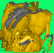
Fig . 3.
The fetus of fig 2. Shown in transparency (volume rendered) mode. This view makes sense when seen rotating. See the associated movie sequences.
Movie 1: Translucent volume rendered representation of this fetus.
Rotating fetus Volume Movie
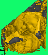
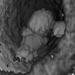
Fig . 4.
A fetal head, displayed with a lower threshold, showing some intra cranial detail, orbits and other bone detail; and lenses of the eyes.
Fig . 5.
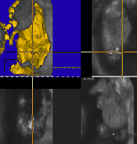
A MPR view of a fetal trunk. This dataset was processed to segment out the bladder. In doing so the urethra was also clearly visualised, from the bladder through the penis, since the fetus happened to be passing urine during the image acquisition. This detail is much more apparent in the associated movie sequence following.
More easily visible in the original image is the orange shaded volume representing anechoic regions.
Movie 3:
Fetal Peeing Movie
Fig . 6.
A very young fetus of about 6 weeks, within the uterine cavity, recorded from a transvaginal scan. The fetal crown-rump length is only 2 cm. At this stage the neural groove is open; the limb buds incompletely formed; the stomach lies in the umbilicus, externally to the abdomen. These details are all clearly seen in this 3D scan. The yolk sac is seen on the uterine wall to the right of the fetus.
 US of Fetal heart
  Fetal MRI studies
  Personnel