| Read Display Info if you have difficulty browsing these graphics-orientated pages.To view a movie, click on an image with a [GIF] or [AVI] or [WMV] link) | |
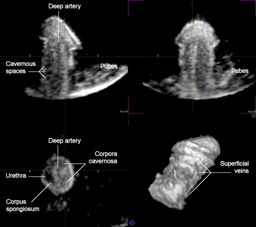
| 3D & 4D US
Click here to see Penile Erection during Real Intercourse [WMV 1.773KB] Click below to see Penile Erection Using an Artificial (Acoustic) Vagina [AVI 776KB]. |
|
 |
|
| A pair
of pictures linked here shows the changes in penile volume and tissue
"density" before and after the use of viagra. These
following links show the pre-
and post- viagra blood flow
change. |
|
| A link here shows the reversed flow
(in blue) through one side of the penile artery, possibly from a
shunting flow (in red) from the other side. |
|
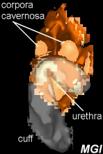
| 3D MRI A front view of the penis and the artificial sphincter (cuff) only about 2/3 around the spongiose urethra [AVI 255KB]. A successfully implanted cuff around the spongiose urethra and the inflatable balloon near the bladder [GIF 356KB]. |
|
 |
 |
