| Read Display Info if you have difficulty browsing these graphics-orientated pages.To view a movie, click on an image with a [GIF] or [AVI] link) | |
| Real-Time
3D Kissing Lips |
|
 |
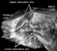 |
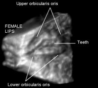
| Above: The left image shows the coronal
view of the orbicularis oris of the female lips without the skin. The
right image also shows the (left lateral) sagittal view of the same
named muscle in the (pouting) male lips. Click on either image above
for a 4D movie (AVI, 2.5MB), or click here for a smaller
movie (WMV, 640KB). Note that the two pairs of lips do not appear in the
same scene in the movie. |
|
| Normal
3D &
4D View How do you pout? |
|
 |
 |
|
Dynamic 3D (4D) a
front view [GIF 90 KB] & a rotational
view
[AVI 472KB] with right-side lip muscles disclosed.
|
|
 |
Functional 2D images reformatted from the same 4D data (upper [GIF 135 KB]). See how the tiny muscles in your lips (two sets of the folding deeper & superficial orbicularis oris) straighten out when you pout. |