| Read Display
Info if you have difficulty browsing these graphics-orientated
pages.To view a movie, click on an image with a [GIF] or [AVI] link) |
|
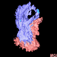
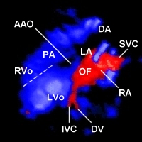
Red & blue in the left image show flows
towards & away from the probe located behind this page. The red indicates
flows from the superior and inferior vena cava (SVC, IVC) returning to
the right atrium (RA), with partial flow through the oval foramen (OF)
into the left atrium (LA). The blues are systolic flows from the right
and left ventricular outlets (RVo, LVo) into the pulmonary trunk (PT) and
ascending aorta (AAO, mostly obscured by the red), correspondingly. The
blues in the two ventricles appear to merge along the dotted line due to
colour smearing artefacts, which are inherited from cross-sectional ultrasound
and need to be overcome. |
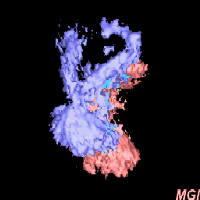
| The 4D intracardiovascular flow from a 32-week fetal
heart is viewed left-inferio-posteriorly. Click here[AVI
77KB] to see a movie of the flow without showing the heart. Click here
[AVI 444KB]to see a movie of the same flow running
through the heart. |
|
|
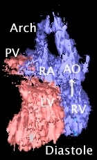
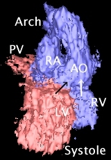
| The unique dynamic
diagnostic potential of the 4D echocardiography for prenatal detection
of the complex cardiac defects. The above two images are reconstructed
from a 4D dataset created using both grey-scale and colour Doppler information
from a 22-week fetus. The structures are coloured according to anatomy
(not flow direction). The movie linked from
the diastolic image [GIF 307KB] shows that both the ascending aorta
(AAO) and pulmonary trunk (PT, behind the AAO) arise exclusively from the
right ventricle (RV, white arrow), i.e., double-outlet right ventricle
(DORV). This was consistent with cross-sectional findings. Because they
are diastolic views when blood flow in the AAO and PT is slow, narrow flow
paths imaged. The movie linked from the systolic
image [GIF 343KB] shows wider paths due to rapid systolic flow. Note
that the shunting flow from the left ventricule (LV) through the ventricular
septal defect (black arrow) reveals the overriding of the aorta, only seen
in systole, with the narrowed PT. The overall diagnosis is then tetralogy
of Fallot, with associated DORV. Arch: aortic arch; PV: pulmonary vein;
RA: right atrium. |