Prenatal Echocardiography in Three and Four Dimensions

Read Display Info if you have difficulty browsing these graphics-orientated pages. To view a movie, click on an image with a [GIF] or [AVI] link)
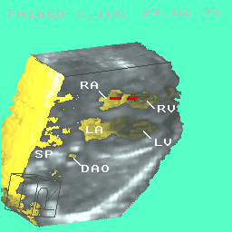
A dynamic 3D (4D) cardiac movie from a 19- week fetus [GIF 744 KB] [AVI 57 KB]. SP: spine. 
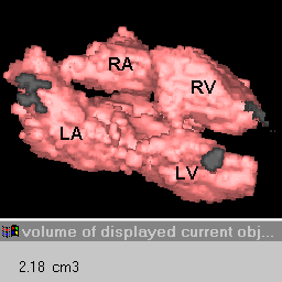
Negative display of the chambers of the same heart. It makes it convenient to measure the cardiac volumes of irregular shapes.
[.gif 38kb]
Interactive multiplanar reformatting of the 3D object [(o)s in upper & lower sets] with different orientations providing unlimited sets of three orthogonal 2D planes using only one scan data set. Only shown here are - Upper: (a) left ventricular short axis view; (b) left heart two-chamber view; and (c) 4-chamber view. Lower: (a) long axis view of left heart, (b) cardiac base short axis view, and (c) bi-ventricular view. The positions of the planes (a), (b) and (c) were indicated by section lines a,b,c. AO: aorta; PA: pulmonary artery; RVOT: right ventricular outflow tract.
[.gif 41kb]
Paper Abstract [Ultrasound in Medicine and Biology 1996; 22: 979-86]
A three-dimensional (3D) acquisition system using an electromagnetic position sensor, attached to a standard transducer on an unmodified ultrasound scanner, was developed to capture two-dimensional (2D) fetal echocardiograms at various positions and orientations. Operating in real-time directed M-mode allowed recording of 2D structural images and cardiac motion curves from which the fetal cardiac phase could be determined. By digitising over a hundred image frames for each scanning sequence, and by selecting frames at the particular phases, 3D views of the fetal heart were reconstructed for each phase. Of 20 sequences of six fetuses scanned, 13 sequences successfully demonstrated usable 3D fetal heart structures, including four cardiac chambers, ventricular and atrial septa, foramen ovale, and some of the cardiac valves and great vessels. Re-arrangement of those phased 3D images into a cyclic sequence could generate dynamic 3D views of a beating fetal heart. We believe that, with further technical development, this new approach will be of use in the diagnosis of prenatal cardiac malformations and malfunction, in in-utero cardiac surgery, and in fetal cardiology teaching.