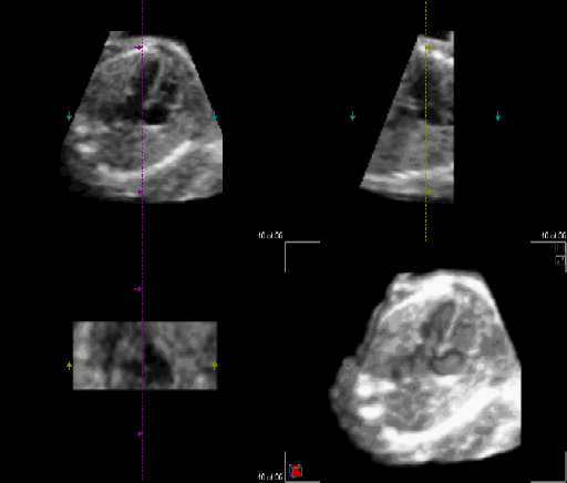
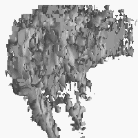
| Click on
the image above
for a dynamic 2D display [GIF 266KB],
reformatted
from a 4D dataset of a 22-week fetal heart, acquired using a Real-Time
Volumetric Ultrasound System. Click here for a 4D
surface display [AVI 479KB] of the same heart, cut open at the
four-chamber-view
level, showing major structures. The normal fine structures, such as
foramen
valve, interatrial and interventricular septa, and the tricuspid and
mitral
valves, are seen over-thickened, both because the spatial resolution is
insufficient and because the structures are perpendicular to the sound
direction. |
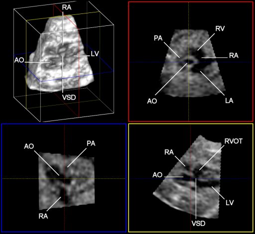
A negative
4D surface
display [GIF 252KB] (i.e., cardiovascular cavities shown as solid),
disclosing the spatial relationships between the major structures of a
26-week fetal heart. Due to insufficient image resolution, fine
structures
cannot be rendered. Click here for a dynamic
2D image [AVI 240KB] and here for a (possitive) 4D
surface display [AVI 454KB] from the same dataset. AAO:
ascending
aorta; AD: arterial duct; Arch: aortic arch; DAO:
descending aorta; ICV: inferior caval vein; PT: pulmonary
trunk;
PV: pulmonary vein; RA & LA: right &
left
atria; RV & LV: right & left ventricles;
RVi & RVo:
right ventricular inlet & outlet; VD: venous duct. |