| Read Display Info if you have difficulty browsing these graphics-orientated pages.To view a movie, click on an image with a [GIF] or [AVI] link) | |
| Normal 3D & 4D View
Why does this upper eyelid have double folds? |
|
 |
 |
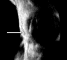
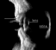
| 2D sagittal views reformatted from a 4D data set, showing the thin dark muscular layer in the upper eyelid bifurcates when the eye opens. See the eye blinking [AVI 129KB]. | |
| Look into & out through your pupils | |
 |
 |
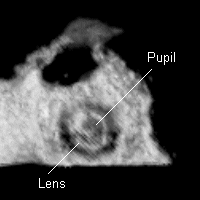
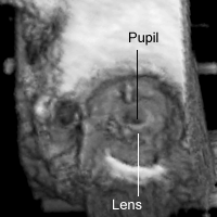
| This 3D image shows how your lens and iris (& pupil) may look like when viewed between your open eye lids [AVI 286KB]. | This 3D image shows how your lens and iris (& pupil) may look like when viewed from your retina [AVI 407KB]. |
| As ultrasound can be performed without any lighting, it is possible to measure the pupillary diameter in the total darkness. But so far, we do not know if this has any clinical significance. | |
